Basal Cell Carcinoma vs Squamous Cell Carcinoma: Understanding the Differences
A skin cancer diagnosis raises immediate questions. If you've been diagnosed with basal cell carcinoma (BCC) or squamous cell carcinoma (SCC), you need clear information about your condition and treatment options. Understanding BCC vs SCC helps you make informed decisions about your care, as these two skin cancers differ in appearance, behaviour, and treatment approaches.
Both BCC and SCC respond well to treatment. Cure rates exceed 90% when detected early [1]. The UK diagnoses approximately 156,000 non-melanoma skin cancers each year [2].

What Are BCC and SCC?
Basal cell carcinoma develops from basal cells in the epidermis's deepest layer. These cells normally divide to replace aging skin cells. UV radiation damages their DNA, causing uncontrolled multiplication. BCC represents 80% of UK non-melanoma skin cancers [3].
Squamous cell carcinoma begins in the flat squamous cells forming your skin's outer layers. These cells sit closer to the surface than basal cells. SCC accounts for 20% of UK non-melanoma skin cancers and behaves more aggressively than BCC, though early treatment remains effective [3].
Visual Appearance: How to Tell Them Apart
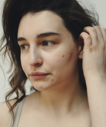
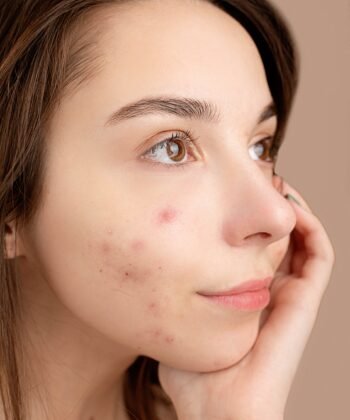
BCC typically presents as a pearly or waxy bump with visible blood vessels. Some BCCs appear flat and scar-like, either flesh-coloured or brown, expanding slowly over months or years. A common sign: a sore that bleeds, forms a scab, seems to heal, then bleeds again. Pink growths with raised, rolled edges and central indentations also indicate BCC.
SCC looks different. You might notice a firm red nodule or a flat lesion with a scaly, crusted surface. The texture often feels rough, like sandpaper catching on clothing. SCCs tend to look inflamed and may resemble persistent eczema that won't respond to standard treatments.
Location Patterns
BCC favours areas receiving intense but intermittent sun exposure. The face (particularly the nose), neck, shoulders, and back see most BCCs. Think of spots that burn during beach holidays but stay covered during daily activities.
SCC develops where skin accumulates decades of sun damage. The backs of hands, lips, ears, and balding scalps host most SCCs. These areas catch daily UV exposure during routine outdoor activities, even in Britain's cloudy weather.
Risk Factors and Causes
UV exposure drives both cancers, but exposure patterns differ. BCC links to intermittent, intense sun exposure. Those childhood sunburns from summer holidays increase BCC risk, especially in fair-skinned people who burn rather than tan.
SCC correlates with cumulative lifetime sun exposure. Outdoor workers, gardeners, and sports enthusiasts show higher SCC rates. Additional SCC risk factors include immunosuppression from medications or medical conditions, previous radiation therapy, chronic wounds or scars, and certain HPV infections.
Growth Rate and Spread Risk
BCC grows slowly, sometimes unnoticed for years. Metastasis remains extremely rare (under 0.1% of cases). However, untreated BCCs can invade local tissues, damaging cartilage or bone [4].
SCC grows faster and spreads in 2-5% of cases [5,6]. Larger tumours, those on lips or ears, and those in immunosuppressed patients carry higher metastatic risk.
Basal Cell Carcinoma (BCC) vs Squamous Cell Carcinoma (SCC)
| Feature | Basal Cell Carcinoma (BCC) | Squamous Cell Carcinoma (SCC) |
|---|---|---|
| Prevalence | Most common type of skin cancer UK: ~80% of non-melanoma skin cancers |
Second most common type UK: ~20% of non-melanoma skin cancers |
| Appearance | • Shiny, pearly or waxy bump • Flat, flesh-colored or brown scar-like lesion • Bleeding or scabbing sore that heals and returns • Pink growth with rolled edges |
• Firm, red nodule • Flat lesion with scaly, crusted surface • New sore or raised area on old scar • Rough, scaly patch that may bleed |
| Common Locations | • Face (especially nose) • Neck • Shoulders • Back |
• Face, lips, and ears • Backs of hands • Scalp • Lower legs |
| Growth Rate | Slow-growing Develops over months to years |
Faster growth than BCC Develops over weeks to months |
| Spread Risk | Very rarely spreads (<0.1%) Stays local to original site |
Small risk of spreading (2-5%) Can reach lymph nodes if untreated |
| Main Risk Factors | • Intermittent intense sun exposure • Childhood sunburns • Fair skin • Age over 50 |
• Cumulative sun exposure • Weakened immune system • Chronic wounds or scars • Previous radiation |
| Treatment Options | • Surgical removal • Mohs surgery for face • Freezing (cryotherapy) • Topical creams • Light therapy |
• Surgical removal with wider margins • Mohs surgery • Radiation therapy • Combined treatments for advanced cases |
| Treatment Success | Excellent outcomes • Cure rate: Over 95% • Most are completely cured |
Very good outcomes • Cure rate: Over 90% when caught early • Regular monitoring important |
| After Treatment | • Skin checks every 6-12 months • Daily sun protection • Watch for new spots |
• Skin checks every 3-6 months initially • Lymph node checks • Close monitoring needed |
Treatment Approaches at The Dermatology Clinic London
Treatment depends on tumour type, size, location, and your overall health. Our Harley Street clinic provides comprehensive treatment options.
Surgical excision remains the gold standard. It offers the highest cure rates and allows complete tissue examination. For facial lesions or recurrent tumours, Mohs micrographic surgery provides 99% cure rates while preserving healthy tissue.
Some superficial BCCs respond to topical treatments like imiquimod or photodynamic therapy. Early SCCs in patients unsuitable for surgery might receive cryotherapy or radiation.
Prognosis and Prevention
Treatment outcomes are excellent. Standard surgical excision achieves BCC cure rates above 95%. Mohs surgery reaches 99% success rates [7,8]. Early-stage SCC treatment succeeds in over 90% of cases [9]. However, one skin cancer increases your risk for others. Between 30-50% of patients develop another skin cancer within five years [10].
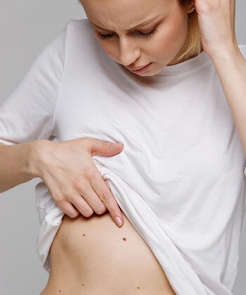
Prevention requires daily sun protection and regular skin monitoring. Professional skin checks every 6-12 months catch new lesions early. Our mole mapping service documents your skin comprehensively, making changes easier to spot.
Don't wait if you notice suspicious skin changes. Early detection significantly improves treatment success and reduces complications.

Medically reviewed by Dr. Daniel Glass
UK trained Consultant Dermatologist Dr Daniel Glass is a General Medical Council registered skin specialist, qualified in both adult and paediatric Dermatology.
REFERENCES
[1] Skin Cancer Foundation. Basal Cell Carcinoma Treatment.
[2] Cancer Research UK. Non-melanoma skin cancer statistics.
[3] British Association of Dermatologists. New data shows a record 224,000 skin cancers in England in 2019.
[4] National Center for Biotechnology Information. Basal Cell Carcinoma. StatPearls.
[5] Caudill J, et al. The risk of metastases from squamous cell carcinoma of the skin. International Journal of Dermatology. 2023;62(4):483-490.
[6] Brantsch KD, et al. Analysis of risk factors determining prognosis of cutaneous squamous-cell carcinoma: a prospective study. Lancet Oncol. 2008;9(8):713-720.
[7] Medscape. Surgical Treatment of Basal Cell Carcinoma.
[8] National Cancer Institute. Skin Cancer Treatment (PDQ®).
[9] Moffitt Cancer Center. Squamous Cell Carcinoma Survival Rate.
[10] Medscape. Basal Cell Carcinoma Treatment & Management.